Adolescent Peri-Acetabular Osteotomy (PAO)
17 year old boy with painful left hip. Note large dysplastic acetabulum and femoral head subluxation on the patient’s left side. The head of the femur is moving in a false socket high on the side of the pelvis. The arthrogram below shows that the hip can be put back into the true socket.


During the arthrogram shown below, radiographic dye is injected into the hip joint. The dark area is fluid that is filling the joint space. In the first image the hip is abducted and internally rotated to demonstrate that the femoral head slides lower into the socket leaving fluid above the hip joint. The second image shows where the femoral head sits when the thigh is in neutral position as it is during standing. This is the position of the hip during the first x-ray above. It was determined that PAO might be possible to place the intact joint surface in a better position to support the femoral head during weight-bearing. The PAO will rotate the socket so that it is more on top of the femoral head.


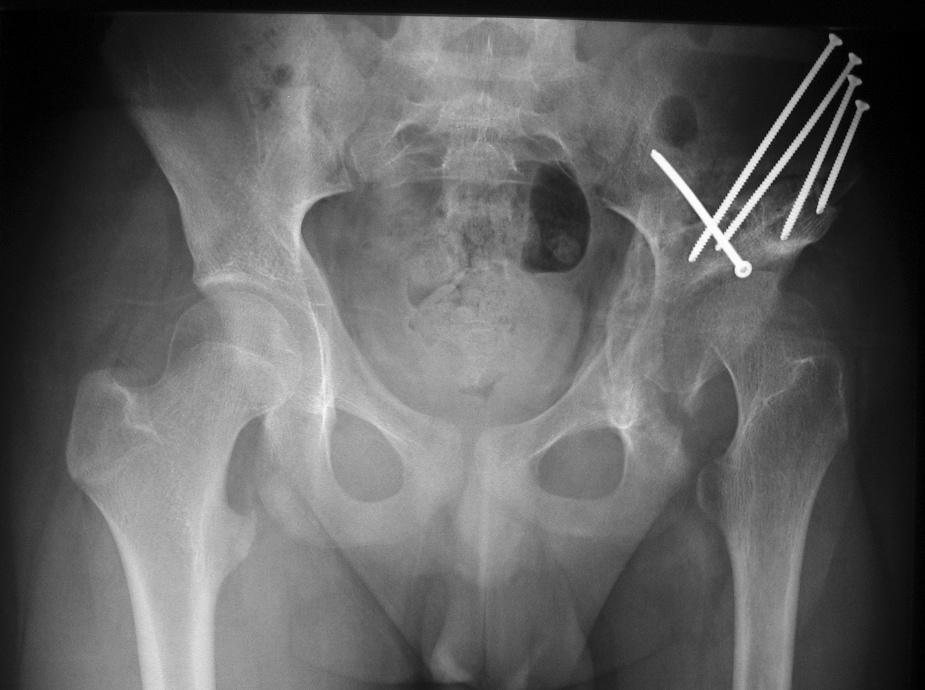
The x-ray below shows the hip after PAO. Note that slope of the socket is more horizontal and the intact portion of the acetabulum has been rotated so that it is directly over the femoral head. This allows the intact cartilage surfaces to support weight-bearing. The patient was relieved of pain and should be able to avoid total joint replacement for many years.