
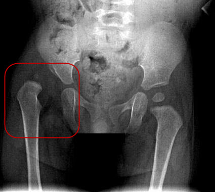
For babies 4 months of age or older and children, x-rays are performed when hip dysplasia is suspected. This is necessary to make the diagnosis or to be sure the hip is normal.
During treatment x-rays can reveal the progress of the hip as it improves. Most children do not need surgery, but for those who do, an arthrogram (x-ray dye injected into the hip joint) at the beginning of the surgery can help the surgeon decide exactly what needs to be corrected.


This child’s left hips is slightly out of the socket. But it goes back into the socket when the legs are held apart. The first principle of treatment is to hold the hip in the joint until it grows and becomes more stable. This can often be achieved with abduction braces or harnesses in infants.


The left hip of this infant is dislocated. It goes into the socket when the hip is held in a flexed position in the Pavlik Harness.
For more information about x-rays, see the Radiology Info website.